Impact: How we developed a new window to the wonders of the living microscopic world
-
Tagged under
Environment and sustainability
-
Lead Academic
Dr Philippe Laissue
Observing live biological samples in real time without disturbing them is one of the great aspirations in the world of microscopy. Dr Philippe Laissue has worked with Cairn Research to develop a new type of microscope - the Large Selective Plane Illuminator (L-SPI) - which allows live samples to be looked at like never before.
The challenge
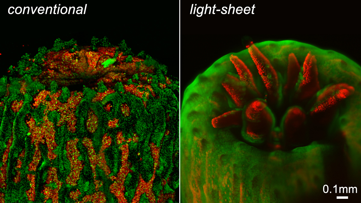
Currently the most effective method of observing a biological process in real time is fluorescence microscopy.
But this process involves light, which can damage the very sample being studied - be it a cell, tissue or organism. This light-induced damage to cells, known as photodamage, means observing without disturbing is very difficult to achieve – and often impossible if the sample is highly sensitive to light.
However, there is a technique that greatly minimises photodamage - light-sheet fluorescence microscopy, also called selective plane illumination microscopy. This technique allows the gentle observation of living things, as it uses far less light than conventional microscopes.
What we did
Dr Laissue’s career has been driven by a fascination for visualising a world invisible to the naked eye. His work focuses on live microscopy using as little light as possible to follow biological events as they unfold with the least possible amount of damage to the observed sample.
In our School of Life Sciences, Dr Laissue had built a first light-sheet prototype to study reef-building corals which are very sensitive to high light. This also makes them the perfect live model organisms to use for testing the effects of microscopy.
Scientific instrument manufacturer Cairn Research wanted to expand its portfolio into the exciting new field of light-sheet microscopy. It successfully applied for an Innovation Voucher from the University of Essex to develop and test a commercial prototype.
The work to create the commercial prototype was conducted at Cairn’s facilities in Kent and in Dr Laissue’s laboratory. Cairn and their highly skilled engineers wanted to produce a prototype unit based on Dr Laissue’s original design and test it extensively, with the aim of validating its user-friendliness and accuracy, and boosting the company’s scientific profile as an innovator.
Funded by a Royal Society Fellowship, Dr Laissue worked with Cairn to develop a completely novel light-sheet illumination device called the L-SPI.
What we changed
Using a unique design approach and philosophy, the L-SPI now offers live-cell imaging for large samples with ultra-low phototoxicity. It is now commercially available and has already gained interest from researchers around the world as it offers long-term 3D imaging of small coral colonies without the observation light stressing the coral.
The L-SPI can be adapted to a wide range of experimental purposes. Its compact size and large freedom of access – such as for positioning micro-electrodes – make it a highly flexible solution. Adding to this flexibility is the fact that it can be fitted to any camera and software.
Dr Laissue said: “I was working with colleagues to get a better insight into coral bleaching and the development of reef-building corals. However, conventional microscopes damage our observed corals, since they use much higher light intensities for the fluorescence. This meant we could observe a coral sample only once, or only for a short period.
“The microscope we developed now means that we can look at coral like never before – very close to the ideal of ‘observing without disturbing’, which really is a microscopist’s dream. We are now getting some fascinating insights into reef-building corals. The microscope also offers great opportunities for looking at other specimens which are as sensitive to light as corals.”