New window to the wonders of the living microscopic world
-
Date
Mon 5 Feb 18
A new microscope which can look at live corals like never before has been developed by Dr Philippe Laissue during his Royal Society Industry Fellowship.
Driven by a fascination for visualising a world invisible to our naked eye, his work on live microscopy uses as little light as possible to follow biological events as they unfold. Less light means less damage to the observed sample – be that a cell, tissue or an organism.
The Fellowship has allowed Dr Laissue, from our School of Life Sciences, to work on a new type of microscopy called light-sheet fluorescence microscopy. This technique allows the gentle observation of living things, as it uses far less light than conventional microscopes.
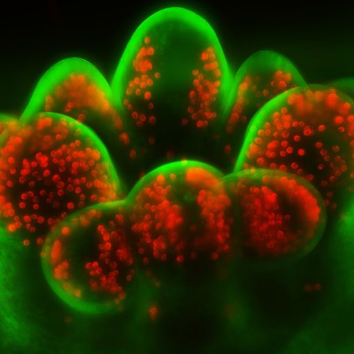
The new microscope, called the Large Selective Plane Illuminator (L-SPI), was developed in collaboration with Cairn Research, an instrument manufacturer in Kent. It is commercially available and has already gained interest from researchers around the world as it offers long-term 3D imaging of small coral colonies without the observation light stressing the coral. This is a major advance, since corals are highly photo-sensitive and easily damaged even by relatively low light levels.
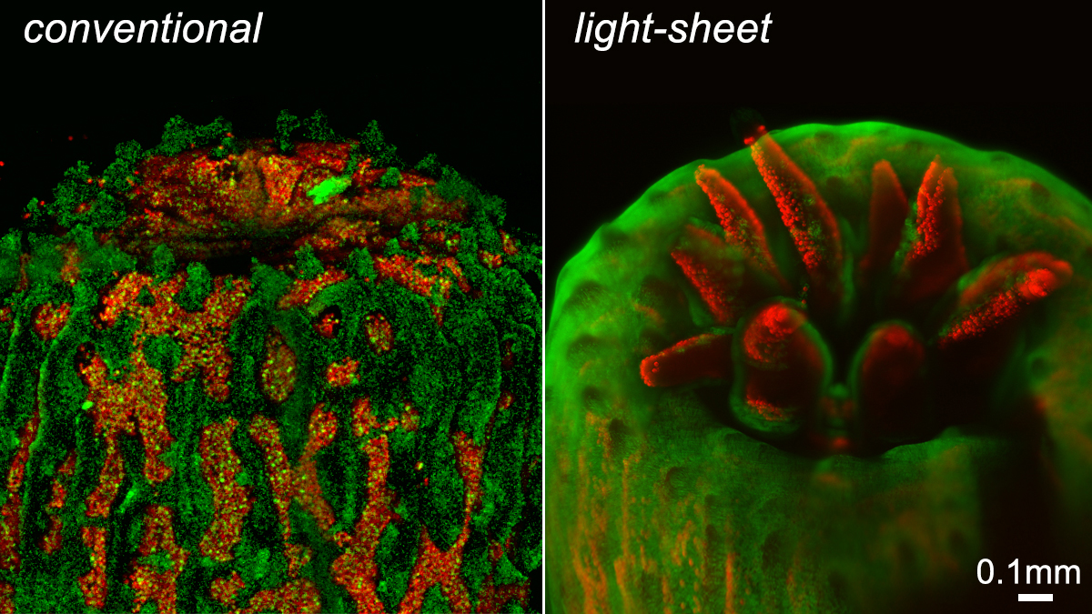
Dr Laissue, who is the Microscopy Facility Manager at Essex, said: “I was working with colleagues in the School’s Coral Reef Research Unit to get a better insight into coral bleaching and the development of reef-building corals. However, it soon became clear that if you use conventional microscopes, they tend to damage the observed coral, since they use much higher light intensities for the fluorescence. This meant we could observe a coral sample only once, or only for a short period.
“The microscope we developed now means that we can look at coral like never before – very close to the ideal of ‘observing without disturbing’, which really is a microscopist’s dream. We are now getting some fascinating insights into reef-building corals. The microscope also offers great opportunities for looking at other specimens which are as sensitive to light as corals.”
He added: “I am very grateful to the Royal Society, which has supported this work from its very modest beginnings. The University of Essex has also been very supportive, showing great flexibility to accommodate this work, which is technically important not just to corals, but to any other live samples, and will further enhance the research being carried out in the School of Biological Sciences.”
Low light microscopy techniques have emerged over the last decade, revealing how easily many organisms and cells can be damaged by light. It is crucial to avoid such damage to ensure that accurate data can be obtained from naturally behaving and developing specimens. Dr Laissue and colleagues have published a paper in Nature Methods which raises awareness of the issue of photo damage, an important consideration for any type of microscopy looking at live organisms.