School of Life Sciences

Our facilities

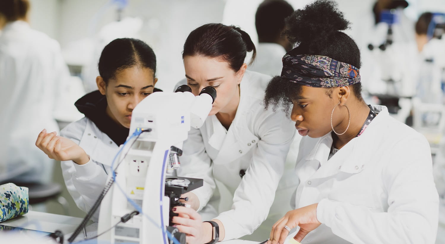
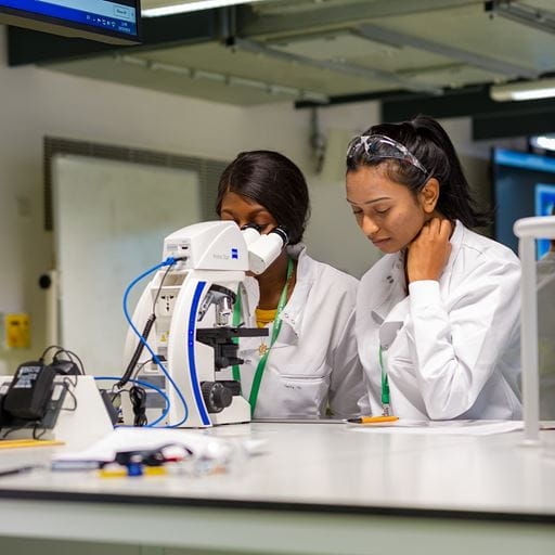
Two students working at a microscope

Cutting-edge environments

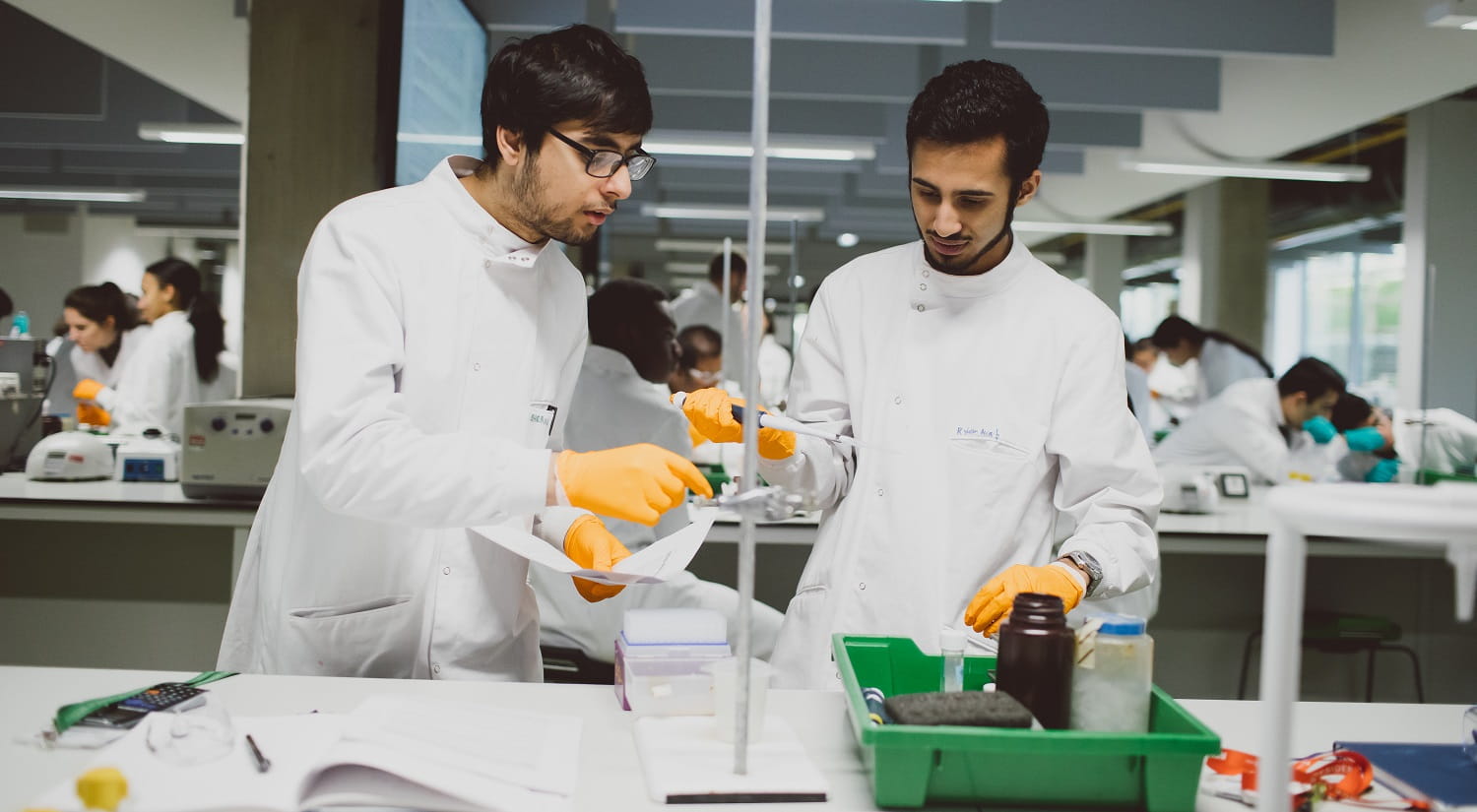
As one of our students, you will be taught in modern laboratories during your practical sessions. These are large and well-equipped, containing up-to-date equipment which gives you valuable experience of using a professional facility.

Final-year and postgraduate research projects are carried out using our academic research laboratories. These were renovated as part of the ongoing expansion of our School and its facilities.

Our aim is to provide you with state-of-the-art equipment and ensure your studies are as relevant and productive as possible.

Examples of our Facilities

STEM Centre

As well as modern breakout rooms, computer facilities and Bytes Cafe, the new STEM Centre is used for many undergraduate practicals and Masters classes.

This enormous laboratory space has new digital microscopes, integrated with the latest audio-visual equipment and displays, as well as Class II microbiological safety cabinets for cell culture, and the latest spectroscopy technology.

The STEM Centre and laboratory spaces are wheelchair accessible, and our lab space has height-adjustable, DDA compliant work benches.

Bioimaging Facility

We have high-end and routine microscopes in darkrooms, workstations for image processing and analysis, and a lab for sample preparation. Using mainly fluorescence microscopy, we are well-equipped to carry out fluorescent protein localisation, morphology and quantified phenotype analysis from single cells to tissues. Areas of expertise are live cell imaging, large-sample macroscopy, and fluorescent-protein-based biosensors.

We also develop our own instruments, algorithms for computational image analysis, and sample preparation techniques. We have a FACS BD Accuri C6 flow cytometer equipped with a blue and red laser, two light scatter detectors, and four fluorescence detectors with optical filters optimised for the detection of fluorochromes, including FITC, PE, PerCP, and APC, and can also detect many variants of fluorescent proteins, such as GFP, YFP, and mCherry.

The School is also in the process of purchasing a new FACS-sorter for separation and isolation of primary human cells and genetically modified cells. The sorter will support applications from all research groups in the School of Life Sciences such as CRISPR/Cas9, purification of pure cancer cell populations for further analysis, advance immunology and isolation of plant cell organelles.
Bioinformatics and Computational Biology

The department now has two 'dry laboratories' and a high-performance computing cluster accessible on and off campus (via VPN), and is configured for both interactive and batch use. The cluster currently has 128 processor cores and a total of 2 Tb of memory, plus 100 Tb of storage, and a separate webserver. Expansion of this system is ongoing.

A broad range of bioinformatics software is installed including all commonly used DNA sequence analysis software and numerous R packages. There is a Galaxy instance on the webserver for teaching purposes. The cluster is in use by numerous postgraduate students, staff and some undergraduate students and actively supported by a dedicated staff member and the genomics group.

Biophysical and Biochemical Facilities

The School is fully equipped to perform cutting-edge biophysical and biochemical research using a combination of conventional and custom designed apparatus including:

  • circular dichroism to monitor secondary structures including chemical and thermal denaturation;
  • stopped flow optical/fluorescence and laser flash photolysis set-ups for kinetic analysis of enzyme reactions and ligand binding;
  • extensive facilities for UV/visible/near infra-red and fluorescence spectroscopy from isolated samples and for proteins within cells (Olis instrument);
  • a microcal isothermal titration calorimetry instrument for deriving the thermodynamics of protein ligand binding;
  • surface plasmon resonance (SPR) facilities to permit the study of a range of biomolecular interactions;
  • extensive protein production and purification facilities;
  • specialist facilities for X-band Electron Paramagnetic Resonance (EPR) spectroscopy – including a liquid helium cryostat and apparatus for preparation of rapid-freeze quench samples;
  • potentiometry set-up for measurement of redox potentials by cyclic voltammetry and redox titration.

Structural bioinformatics /computational chemistry facilities for modelling protein structures include specialist drug design software (from Schrödinger) and high performance GPU-based computing designed specifically for molecular dynamics simulations.

A crystallisation laboratory is equipped with a state of the art ‘Gryphon’ crystallisation robot, Leica M125 microscopes (with digital camera) for crystal viewing, variable-temperature vibration-free crystal incubators, full facilities for ‘manual’ crystallisation, and apparatus for manipulating, cryo-cooling and transporting protein crystals to synchrotron facilities. A single crystal microspectrophotometer is also in place.

Ecology and Environmental Microbiology Facilities

Field-to-mesocosms sampling

Fieldwork sites are accessed by two fieldwork vehicles with full off-road 4x4 capability, and two (7m & 3m) Rigid Inflatable Boats (RIBS).

Field sampling is facilitated by:

  • Van Veen grab samplers
  • diving PAMs
  • underwater video cameras
  • full face diving masks with communications equipment
  • GPS units
  • multiple remote sensing drones (e.g. Phantoms)
  • field portable spectrophotometers
  • fish surveying equipment including fyke nets, passive traps, pelagic trawls, and backpack electrofishing gear (E-fish 500W)
  • a FluoroProbe profiler for the analysis of chlorophyll with algae class determination.

Experimental work is support through a range of aquatic and terrestrial mesocosms, including:

  • 62 large (>100L) temperature-controlled aquaria for simulating tropical or temperate and fresh or marine systems; replicated environmental outdoor mesocosms simulating tidal and terrestrial systems;
  • a state-of-the-art tropical coral-reef research facility devoted to fully climate-controllable experiments;
  • multiple pH-stating systems to control CO2 concentration for ocean acidification experiments.

Microbial culturing facilities support our experimental work and comprise dozens of constant-temperature (and light) growth rooms, and walk-in incubators, across a -12°C to +40°C gradient, used for culturing both Prokaryotes and Eukaryotes. 

Analytical equipment

All field sampling and experimental work is underpinned by high-accuracy analytical measurements provided by:

  • four gas chromotographs (GCs) with diverse detectors (MS, FID, FPD, ECD) for trace gases
  • hydrocarbon and polysaccharide analyses
  • purpose-built purge-and-trap systems for the cryogenic enrichment of trace gases (one for sulphur gases, one for non-methane hydrocarbons)
  • a Fast Isoprene Sensor; Pfeiffer model QMG 422 mass spectrometer with membrane inlet for measuring dissolved gases
  • a uPLC; a new Nutrient Autoanalysis suite as well as sediment oxygen/nutrient flux systems
  • Dionex anion and cation exchange chromatography
  • an HPLC; atomic absorption spectrophotometer
  • 2 X CTG FastTracka II Fast Repetition Rate Fluorometers with Fast Act Laboratory Systems
  • PAM fluorometers; bench top spectrofluorometers
  • LED Flash-Yield systems
  • Leica DMI600B inverted light Limnology microscope – auto XYZ image capture (including Dark Phase)
  • Gilson liquid-handling robot for high-throughput microbial culturing in microtiter plates
  • a multi-mode (fluorescence, optical density, luminescence) microtiter plate reader.

Molecular ecology

In the post-genomics era, we have invested heavily in high-throughput molecular ecology facilities, based around advances in 2nd and 3rd generation sequencing technologies, which we apply across taxonomic domains and level of biological organisation, these include:

  • Hamilton Robotics liquid handling robot
  • DNA, RNA and protein extraction equipment
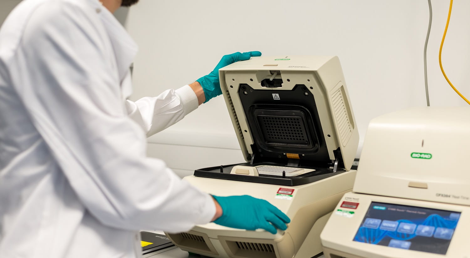
  • banks (dozens) of PCR thermal cyclers
  • a range of 96 and 384 well qPCR machines
  • Blue Pippin DNA size-selection system
  • Stable Isotope Probing equipment and a designated radio isotope laboratory
  • Agilent Bioanalyser
  • NanoDrop 3300 fluorospectrometer and a NanoDrop 1000 spectrometer
  • FLUOstar Omega Microplate Reader
  • an Illumina MiSeq Benchtop Sequencing System. 

Find out more about our ecology and environmental microbiology research group.

Molecular Cell Biology and Cell Culture

The School has excellent facilities for cellular molecular biology research, with dedicated molecular biology and cell/tissue culture rooms facilitating the use of recombinant DNA, the study of protein function, siRNA library screens, various cellular assays and the generation and use of recombinant viruses.

Facilities include:

  • level I and II cell/tissue culture facilities with multiple laminar flow cabinets
  • CO2 incubators
  • FACS and cell sorter facility
  • microscopes
  • centrifuges and ultracentrifuges
  • Bioruptor Sonicator
  • immunohistochemistry suite
  • equipment for mammalian cell manipulation including electroporators and viral transfection technology
  • PCR thermocyclers including quantitative PCR using Taqman and SYBR Green-based methods
  • bioanalyzer
  • Optical/fluorescence/luminescence/ELISA plate readers
  • chemiluminescent, UV and LI-COR imaging systems
  • Illumina MiSeq sequencing machine
  • Metrocubo GPU workstations for compute-intensive molecular dynamics simulations
  • recently refurbished radioisotope suite.

Plant Physiology and Phenotyping

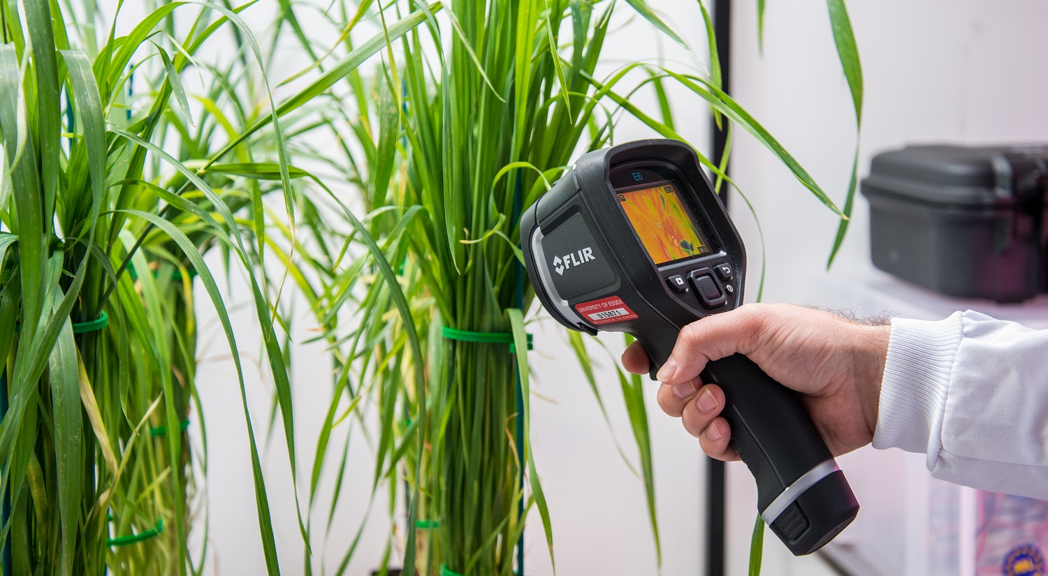
The Plant Productivity Group employs a range of non-invasive techniques including state-of-the-art imaging for plant phenotyping and we have developed and built in-house instrumentation and software to extend our considerable plant physiology suite.

We are fully equipped to perform cutting edge plant physiology and phenotyping research using a combination of conventional and custom designed apparatus including:

  • Chlorophyll fluorescence imaging of whole plants (low res ) and cells (high res)
  • High resolutions imaging of wavelength specific bands (e.g. for imaging ROS)
  • Thermal imaging of stomatal conductance
  • Imaging plant Water Use Efficiency
  • Infra-red gas exchange analysis – including whole plant chambers for Arabidopsis
  • Phenotyping plant form with watering control capabilities and dynamic light regimes
  • Dynamic lighting platforms
  • Spectral reflectance
  • Water regimes including hydraulic conductance.

These state-of-the-art facilities are underpinned by our plant growth capability using computer controlled glasshouse and controlled environment facilities.

Proteomics

The School is well equipped with mass spectrometers including a state-of-the-art hybrid high-resolution LTQ/Orbitrap Velos instrument interfaced with a splitless nanoscale liquid chromatography system for fully automatic genome-scale analysis of protein abundance and post-translational modifications.

Academic and technical staff have extensive experience in quantitative proteomics utilising stable isotope labelling and label-free approaches, mass spectrometry-based analysis of protein phosphorylation and protein-protein interactions.

Smart Technology Experimental Plant Suite

The new Wolfson STEPS is a unique £3.5m facility that is state-of-the-art plant growth facility with cutting edge phenotyping technologies.  Included within STEPS are:

  • A commercially standard vertical farm,
  • An indoor field that replicates real environments anywhere on the globe,
  • Future climate suites that imitate a warming world – with control of CO2 concentration and temperature
  • A ‘crop per drop’ irrigation room for future water that will precisely control water supply and monitor crop water use. 

STEPS has provided the opportunity to extend the existing expertise and facilities in plant phenotyping with the inclusion of a Planteye 3D scanner capable of high-throughout monitoring of plant growth and physiology.

BBSRC Alert22 funding extended capabilities with the development of Dynamic Plant Phenotyping for future proofing crop productivity (DynSCREEN) which includes a PSI dynamic XYZ robotic plant phenotyping gantry system is housed within the dynamic field, and includes thermal, chlorophyll fluorescence, RGB and hyperspectral cameras, leading the way in UK phenotyping. 

Phenotyping in STEPS

STEPS is home to several state-of-the-art phenotyping capabilities including;

  • PlantEye F600 – a multispectral 3D scanner for non-destructive growth measurements and high-throughput scanning of morphological and spectral parameters.
  • DynSCREEN – includes a XYZ robotic system for scanning the indoor field for the following: Hyperspectral (400-2500 nm); Thermal imaging; Chlorophyll fluorescence imaging.

BBSRC ALERT Funded Equipment

We are very pleased to have a number of pieces of equipment funded via the Biotechnology and Biological Sciences Research Council (BBSRC) on behalf of UK Research and Innovation.

The equipment listed below is accessible to academic, public, and private sector users to support development of high-quality research. For more information or to enquire about usage please contact sls-res-manager@essex.ac.uk

EPR Spectrometer – the key machine in our Biomedical EPR Facility

Funded from the BBSRC ALERT award, the Bruker E500 CW X-band EPR spectrometer is used by many national and international users as part of our Biomedical EPR Facility.

This equipment allows for detecting paramagnetic states of biologically significant molecules, such as, proteins, enzymes, and many other molecules, whether small or large, isolated or in complex environments such as cells or tissues. The measurements are possible at both low and room temperatures, including solidly frozen samples at as low temperatures as 4 K, as well as of liquid samples at room temperatures.

Lightsheet Microscope

In collaboration with Eastern Arc Academic Research Consortium (EARC), a grant was awarded for a state-of-the-art lightsheet microscope which enables scientists to observe delicate cells, tissues and whole organisms without damaging them with intense light. This is used in combination with cutting-edge software to address new and exciting biological questions in biomedical and environmental research. It forms part of the Imaging Platform Alliance (IPA) between the EARC universities.

Use of IPA equipment can be applied for through EARC.

Dynamic Plant Phenotyping for future proofing crop productivity (DynSCREEN)

BBSRC ALERT funding has enabled the development of a state-of-the-art phenotyping platform DynSCREEN for Dynamic Plant Phenotyping for future proofing crop productivity. This houses a PSI Plantscreen™ Robotic XYZ System which enables high-precision automated phenotyping of plants for a range of sizes. It includes a range of specialised cameras which can capture spatial and temporal images of plants under various conditions and use these to determine key traits.

This system is part of our Smart Technology Experimental Plant Suite (STEPS), a novel dynamic indoor environment, capable of mimicking any outdoor environment both now and in the future DynSCREEN represents critical investment for UK plant phenotyping and is aimed at promoting collaboration.

Dianthus (NanoTemper)

Dianthus (NanoTemper) is the first ever screening platform to use two biophysical modalities, Temperature Related Intensity Change (TRIC) and Spectral Shift (SS), to measure the strength of molecular interactions (TRICSS).

It has unique features that allow for increased sensitivity in the detection of true molecular interactions:

  • measures affinities between a wide range of fluorescent labelled targets (protein, peptide antibody, DNA, RNA and aptamer) and an even wider range of ligands (protein, peptide antibody, DNA, RNA, aptamer, compounds, fragments, ions, virus like or other particles).
  • generates a significantly lower percentage of false positive results compared to other biophysical techniques and detects a wide range of physiological binding affinities (pM to mM).
  • no immobilisation is required (overcoming Biolayer Interferometry (BLI) and Surface Plasmon Resonance (SPR) limitations), and it differentiates between binding and aggregation (overcoming Isothermal Titration Calorimetry (ITC) limitations).
  • All measurements are in solution and mass-independent and therefore ideal for fragment library screening.
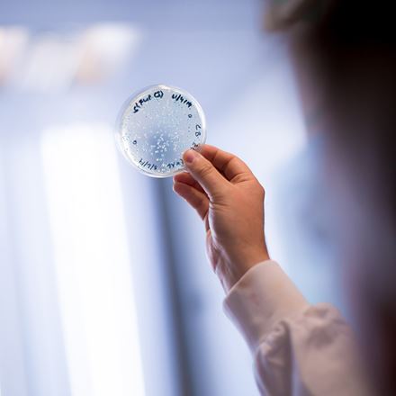
A sample being held up to the light in a lab
Our research

Our research covers the whole spectrum of biology – from the cell right through to communities and ecosystems. Our dedicated research groups bring together experts for collaboration research projects that deliver real-world impact.

See more
Get in touch
David Knight Technical Services Enquiries
Gregor Grant Technical Services Enquiries